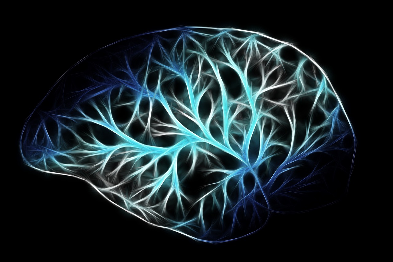
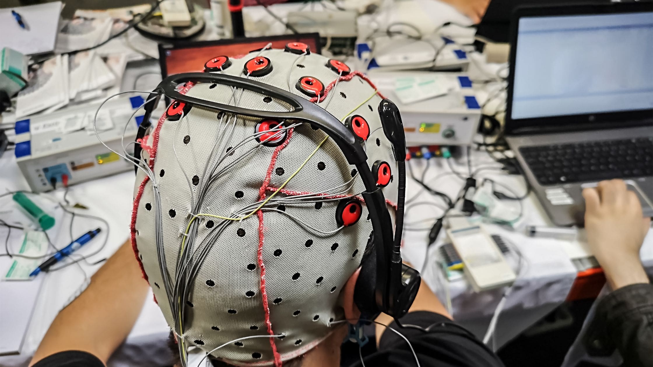
Brain mapping is a technique used to identify various parts of the brain and discover how they all work together. Given the many complexities of the human brain, brain mapping itself is rather complex. The average human brain contains about 86 billion neurons (or nerve cells). When these neurons link up, thousands of different paths could be created, many of which we do not yet understand and therefore can’t predict.
Therefore, brain mapping helps scientists accomplish many goals. For example, it helps them develop a better understanding of the anatomy of the brain and how humans learn. Additionally, it helps doctors to learn more about brain injuries and other brain-related conditions—such as Alzheimer’s, autism, and Parkinson’s. This may lead to more effective treatments and earlier identification. It can also help brain surgeons.
By developing brain maps, scientists can also develop greater insight into the brain’s activity based on different variables. For example, scientists will use brain maps to identify which parts of the brain are responsible for laughter or curiosity; panic and anxiety; joy or depression, and a range of other emotive responses. Here’s what you need to know about the past, present, and future of brain mapping:
Talairach Coordinates
A number of mapping techniques—also referred to as coordinate systems, or atlases—have been developed over time. When developing these systems, scientists build upon previous studies and harness the use of technological advances to create more detailed brain maps.
The first major brain map technique to be popularly used was called Talairach Coordinates. It used a three-dimensional coordinate system to map the location of brain structures regardless of the size and shape of the individual brain. Originally released in 1988, the Talairach method is based on making two anchors, the anterior commissure and the posterior commissure, lie on a straight horizontal line.
By using these coordinates as a basis for identifying anatomical landmarks, it became easier to spatially interpret a brain image through MRI and PET scans. Talairach Coordinates used Brodmann areas—the color-coded mapped illustration identifying the 43 different parts of the cerebral cortex—as guidelines for this mapping technique.
However, Talairach Coordinates have largely since been replaced by the MNI (Montreal Neurological Institute and Hospital) coordinate system, which is the template used for statistical parametric mapping (SPM). SPM is the technique used to examine differences in brain activity recorded during functional neuroimaging experiments.
MAP-sequencing (MAPseq)
A more modern brain mapping technique involves the use of pigmented proteins and microscopes to map neuronal connections. By using the color of a particular protein with a certain region or function of the brain, scientists are able to discern what a patient is doing or thinking when that color appears under a microscope.
While this mapping technique has brought about a major breakthrough in better identifying certain brain functions and neuron paths, it remains a slow, tedious, and expensive method. A new technique called MAP-sequencing, commonly called MAPseq, revolutionized brain mapping just a few short years ago.
This technique uses a giant library of viruses that contain random RNA (Ribonucleic acid) sequences. The library, or database, is then injected into the brain where one virus enters one neuron, thus providing each neuron its own RNA barcode. A DNA sequencer is then able to read each barcode and create a matrix that shows how every neuron is mapped to each other. MAPseq was therefore the most advanced and detailed mapping technique to ever be released.
BARseq
MAPseq has since been replaced with BARseq, the “next generation” of MAPseq. The technology used in BARseq builds upon the techniques used in MAPseq to more accurately pinpoint the location of a neuron. Therefore, BARseq is able to not only identify a neuron’s connections (which is what MAPseq does), but also determine patterns of psychological activity and gene expression.
Another problem that arose with MAPseq was that the method’s process of tagging neurons resulted in low special resolution. This led to the creation of blurry maps that made it difficult for scientists to clearly see how a neuron was connected to a particular pillar in the brain. BARseq addresses this issue by tagging and locating neurons in their original form and location in the brain. By doing so, BARseq can provide important information for studying how neural circuits are formed. This may help researchers better understand of brain activity and identify when and how certain brain diseases are formed.
“If we could have a complete wiring diagram, a map, that would provide a foundation for understanding thought, consciousness, decision-making and how those go awry in neuropsychiatric disorders like autism, schizophrenia, depression, OCD,” said Professor Anthony Zador, a neuroscientist based at Cold Spring Harbor Laboratory. His lab, led by postdoctoral fellow Xiaoyin Chen, created BARseq.
The Future of Brain Mapping
While BARseq can accurately locate where a neuron connection begins, at present it can only estimate where it ends. Researchers and scientists are working on increasing spatial resolution at the destination to further improve BARseq’s effectiveness.
MAPseq, BARseq is a cheaper, faster, and more advanced brain mapping technique than those that came before it. There is still work to be done to further refine the method and create more accurate results. However, it is the most advanced brain mapping technique ever created. It will play an invaluable role in helping scientists better understand the complexities of the human brain.


